
客户文献解读:PDCD5调控细胞增殖、细胞周期及细胞凋亡
信息来源:k8.com 作者:genecreate 发布时间:2018-04-12 10:16:32
题目:PDCD5 regulates cell proliferation, cell cycle progression and apoptosis
期刊:Oncology letters
影响因子:1.6
研究背景
PDCD5(Programmed cell death 5)是程序性细胞死亡蛋白家族成员,同肿瘤细胞生长及凋亡密切相关。为了研究PDCD5的分子及细胞功能,本文通过慢病毒载体构建了稳定过表达PDCD5的人表皮癌细胞A431细胞株,检测了该细胞株的细胞增殖、细胞周期、细胞凋亡,并进一步验证了PDCD5在肿瘤生长过程中是否通过p53行使相关功能。本研究说明了PDCD5在肿瘤发生过程中的分子功能,PDCD5作为肿瘤抑制因子,在肿瘤治疗中可能成为潜在靶分子。
研究内容及结果
1. 慢病毒包装及过表达稳转株构建
本文所使用的PDCD5过表达慢病毒及稳定过表达PDCD5的A431细胞株订购自武汉k8.com生物工程有限公司(GeneCreate Biological Engineering Co.,Ltd)。通过WB及荧光定量PCR实验证明PDCD5在A431细胞株中显著过表达(Fig.1 A.B.C)。
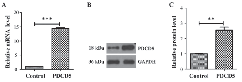
图1 PDCD5过表达水平检测
2. MTT细胞增殖实验
以空载体感染细胞为对照组、以PDCD5过表达A431细胞为实验组进行MTT实验,分别在12h、24h、48h、72h监测数据,绘制生长曲线,可见在72-96h时PDCD5过表达组的增殖显著受到抑制(Fig.2)。
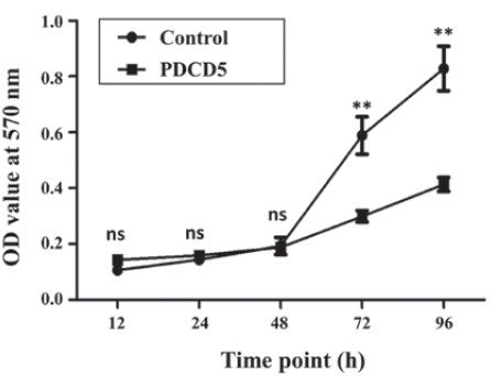
图2 A431细胞增殖
3. 流式细胞术检测细胞周期及凋亡
为了进一步研究PDCD5对于癌细胞增殖抑制的机制,利用PI染色后进行流式细胞术分析,结果显示PDCD5过表达的A431细胞出现G2/M周期阻滞(Fig.3 A.B)。
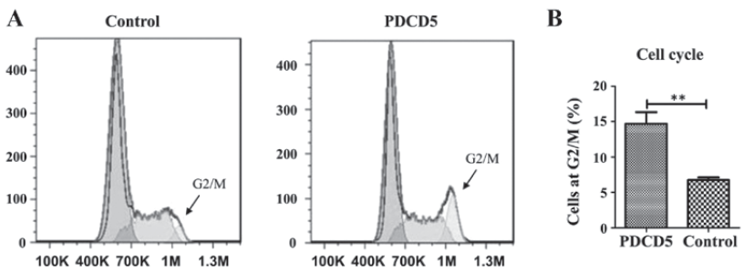
图3 A431细胞周期
为了考察PDCD5对于肿瘤细胞凋亡的影响,使用Annexin V/PI染色及流式细胞术检测过表达PDCD5的A431细胞凋亡,显示PDCD5过表达组凋亡比例显著升高(Fig.4 A.B)。
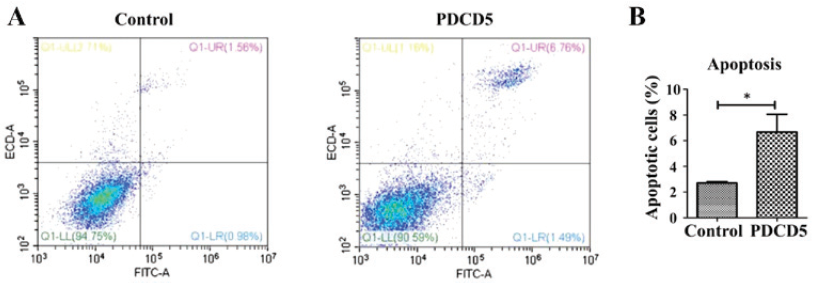
图4 A431细胞凋亡
4. WB及荧光定量PCR检测PDCD5、P53及细胞凋亡相关通路蛋白表达水平
为了进一步研究PDCD5对于肿瘤细胞增殖抑制、周期阻滞的内在机制,通过WB和荧光素定量PCR检测p53、bax、bcl-2、casp3在蛋白和mRNA水平的表达,发现p53、bax和casp3表达升高,bcl-2表达降低(Fig.5 A.B.C)。
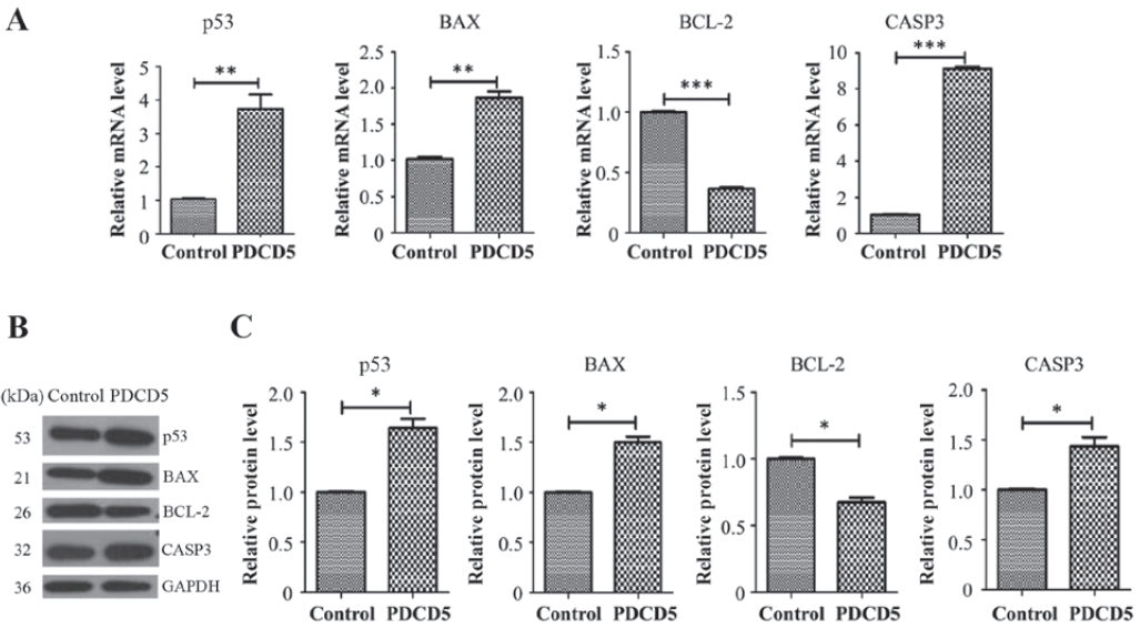
图5 PDCD5过表达细胞中p53、bax、bcl-2、casp3表达水平
文章小结
本文利用慢病毒构建的PDCD5稳定过表达A431细胞株为实验模型,发现PDCD5过表达细胞体增殖受到抑制、细胞周期于G2/M期阻滞、细胞凋亡率增高。进一步研究证明PDCD5过表达细胞中p53表达水平升高,凋亡通路相关基因bax和casp3呈现过表达、bcl-2基因表达降低。PDCD5可能通过经典凋亡信号通路机制影响肿瘤的生长。
解析文献
Penghui Li et al. PDCD5 regulates cell proliferation, cell cycle progression and apoptosis. Oncology letters,2017,15: 1177-1183
参考文献
1. Hipfner DR and Cohen SM: Connecting proliferation and apoptosis in development and disease. Nat Rev Mol Cell Biol, 2004, 5: 805 815.
2. Löbrich M and Jeggo PA: The impact of a negligent G2/M checkpoint on genomic instability and cancer induction. Nat Rev Cancer, 2007, 7: 861 869.
3. Alenzi FQ: Links between apoptosis, proliferation and the cell cycle. Br J Biomed Sci, 2004, 61: 99 102.
4. Liu H, Wang Y, et al: TFAR19, a novel apoptosis related gene cloned from human leukemia cell line TF 1, could enhance apoptosis of some tumor cells induced by growth factor withdrawal. Biochem Biophys Res Commun, 1999, 254: 203 210.
5. Wang N, Lu HS, et al: Involvement of PDCD5 in the regulation of apoptosis in fibroblast like synoviocytes of rheumatoid arthritis. Apoptosis, 2007, 12: 1433 1441.
上一条:多克隆抗体制备流程
下一条:外泌体提取方法有哪些
最新动态
-
06.18
亚细胞定位预测与验证结果不一致的原因是什么?
-
06.18
无需抗体、跨物种通用:三篇高水平论文实证DAP-seq如何助力植物转录调控?
-
06.11
打破“单向用药”的局限!IF=11.9《Asian J Pharm Sci》证实:红参外泌体,口服就能双向调控骨代谢
-
06.11
植物外泌体研究前沿:2026年4—5月6项突破性成果系统梳理
-
06.11
IF 26.8!颠覆“越小越好”的认知:南方医科大团队用40微米的柠檬胶囊,让肠屏障主动“开门”抗癌
-
06.11
从零开始研究一个基因,这篇讲透了!
-
06.11
解码RNA互作奥秘:k8.comRIP/RNA pull-down试剂盒助力多篇高质量研究,深入解析肿瘤及椎间盘退变调控机制
-
06.11
别再大海捞针了!虚拟筛选正在改变药物发现的方式!
-
06.11
BiFC还是LCA?你的蛋白互作该用哪种荧光互补技术?
-
06.11
荧光素酶5大高分案例+实验小技巧!
 X
X